theguardian.com
Cure8 news brief
Cure8 news brief
Better imaging of intestinal cells could help researchers find previously unseen changes in the gut barrier that contribute to IBD, which might eventually guide more targeted therapies or diagnostics.
Researchers and clinicians studying IBD biology; patients and caregivers interested in long-term research directions; funders and science-policy observers.
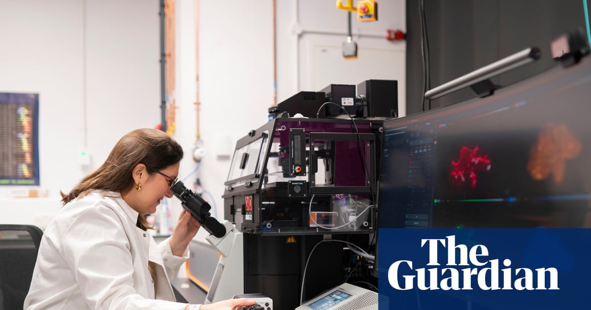
Researchers at the Rosalind Franklin Institute have installed a specialized STED super-resolution microscope (“Curie”) that can image cellular structures down to ~20 nm and study intact tissues and mini-organs at near-physiological temperatures.
The team is using it to visualise protein complexes in the intestinal epithelial barrier across development and in disease, aiming to identify structural differences that could point to root causes of IBD.
This article reports on the instrument, its technical abilities (deformable mirrors, temperature-controlled stage) and early lab use, not on new patient treatments or clinical results. It also notes related UK funding announcements for the institute and wider tensions in research budgets.
What this means: the work is at a laboratory/technology stage focused on basic science and cellular imaging; any implications for new IBD treatments are prospective and would require further research and clinical validation.
This is a technology/early-research news story from a mainstream outlet describing capabilities and research aims rather than reporting clinical findings. Any future therapeutic implications remain speculative until peer-reviewed studies show specific disease mechanisms or validated biomarkers.
Review the original publication for the complete reporting, methods, and context.
This Cure8 brief is based on source text from the linked article. Cure8 is informational only and is not a substitute for professional medical advice, diagnosis, or treatment.
The finding links a specific mucus-associated protein (HMGB1) to barrier defects in Crohn’s disease, which could help explain how bacteria reach and inflame tissue in active lesions and point to future biomarker or...
Researchers report links between copper-driven cell death pathways and IBD biology; identifying cuproptosis-related biomarkers or drugs could open new diagnostic or therapeutic avenues for people with Crohn's disease or...
Researchers are investigating circRNAs as potential biomarkers and therapeutic targets in IBD because they may help explain disease mechanisms and lead to better diagnostics or treatments in the future.

