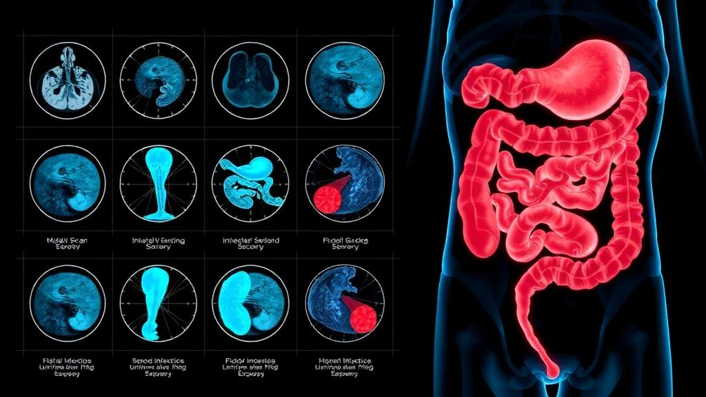
Fusing blood tests, CT enterography radiomics, and deep-learning analysis of deep small-bowel endoscopy images may improve detection of active small-bowel Crohn’s disease — a common clinical challenge because many affected segments lie beyond standard colonoscopy reach.
Who Should Pay Attention
Clinicians who manage Crohn’s disease (especially small-bowel disease), gastroenterology researchers working on imaging and AI, and patients interested in advances in disease activity monitoring.
Study Snapshot
Story typeResearch news
Evidence typeClinical study
Source depthFull source text
What To Know
Researchers used retrospective clinical data and three imaging-related streams: selected laboratory and clinical variables (including CRP and fecal calprotectin), quantitative radiomics from CT enterography, and features plus a binary output from a ResNet-18 model trained on double-balloon enteroscopy images.
They compared single-modality models to combined models and found the fully integrated models performed best by AUC and multiple comparative tests (DeLong, NRI, IDI), and they packaged results as nomograms and used SHAP to explore feature contributions.
How this might be used The approach is primarily a diagnostic/monitoring tool idea: combining systemic biomarkers, cross-sectional imaging, and mucosal images to improve detection of small-bowel activity. The study is retrospective and from a single center; the models would need external validation and prospective testing before clinical use.
Keep In Mind
This is a retrospective, single-center study using double-balloon enteroscopy images and CT enterography radiomics. Findings are promising but require external validation and prospective testing before changing clinical practice. The article summarizes model-building and internal performance metrics rather than reporting clinical outcomes from prospective use.
Source Details
Review the original publication for the complete reporting, methods, and context.
This Cure8 brief is based on source text from the linked article. Cure8 is informational only and is not a substitute for professional medical advice, diagnosis, or treatment.
Understanding how individual diet and microbiome patterns relate to disease activity could help create personalized dietary approaches for children with Crohn's disease and improve monitoring using home fecal...
This model aims to identify healthy first‑degree relatives at higher risk of developing Crohn’s disease before symptoms start, which could enable earlier monitoring or preventive strategies. The score uses simple...