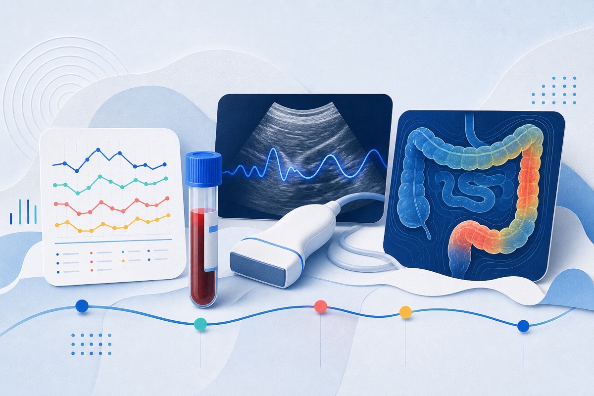
Imaging can reveal characteristic bowel and vascular patterns that help distinguish Crohn's, UC, celiac disease, and systemic autoimmune or vasculitic causes of GI symptoms, which may speed diagnosis and detect dangerous complications early.
Who Should Pay Attention
Clinicians (radiologists, gastroenterologists), researchers studying autoimmune GI disease, and patients with IBD or systemic autoimmune conditions who are undergoing imaging for unexplained GI symptoms.
Study Snapshot
Story typeResearch paper
Evidence typeResearch paper
Source depthJournal abstract
What To Know
The article groups autoimmune GI involvement into three categories: primary GI autoimmune diseases (celiac disease, Crohn's disease, ulcerative colitis); systemic autoimmune diseases with secondary GI involvement (for example systemic sclerosis, lupus, rheumatoid arthritis, Sjögren's); and other autoimmune or vasculitic conditions that can sometimes affect
the gut (for example polyarteritis nodosa, IgA vasculitis, Behçet disease, Takayasu arteritis, dermatomyositis, IgG4-related disease).
The review highlights advances in cross-sectional imaging that allow recognition of mural, mesenteric, and vascular imaging signatures tied to underlying pathophysiology, and stresses combining imaging with clinical and serologic information to improve diagnostic confidence and identify complications such as ischemia, perforation, obstruction, or toxic megacolon.
Keep In Mind
This is an abstract-summary of a narrative review in Abdominal Radiology. It synthesizes imaging patterns rather than reporting new primary data; recommendations are interpretive and should be applied in the context of clinical and serologic information.
Source Details
Review the original publication for the complete reporting, methods, and context.
This Cure8 brief is based on source text from the linked article. Cure8 is informational only and is not a substitute for professional medical advice, diagnosis, or treatment.
Digestive and liver disease : official journal of the Italian Society of Gastroenterology and the Italian Association for the Study of the Liver· via PubMed
IBD may involve small-vessel changes beyond the gut. If reproducible, retinal microvascular alterations detectable by OCTA could offer a non-invasive window into systemic microvascular involvement in IBD.
SpA is a frequent but underrecognized extraintestinal manifestation in people with IBD; validated screening tools can help identify patients who need rheumatology evaluation. Earlier detection may address joint symptoms...