A quantitative, biopsy-based measure of collagen architecture could help study intestinal fibrosis, which underlies stricture formation in Crohn’s disease. Feasibility data support further validation work that may lead to biomarkers linked to disease progression or treatment response.
Who Should Pay Attention
Researchers studying fibrosis and histologic biomarkers; gastrointestinal pathologists and histotechnologists; clinicians and investigators focused on Crohn’s disease complications (strictures) and translational IBD research.
Study Snapshot
Story typeResearch paper
Evidence typeResearch paper
Source depthJournal abstract
What To Know
The researchers applied second harmonic generation/two-photon excitation imaging and qFibrosis morphometry to 31 biopsy samples from 20 patients with Crohn’s disease and annotated compartments (glands/crypts, lamina propria, muscularis mucosae, submucosa).
They report depth-dependent differences: higher overall collagen burden in deeper layers (submucosa) but more dispersed fiber/string architectures in superficial compartments. The paper frames these results as feasible and preliminary rather than clinically validated.
This was a pilot feasibility study without non-IBD control tissues or paired histochemical stains (Sirius Red/trichrome) for validation, so the findings should be seen as methodological and hypothesis-generating—useful for researchers planning larger validation cohorts.
Keep In Mind
This report is a pilot feasibility study (abstract-level/full-text) using routine FFPE biopsies but lacks non-IBD controls and paired histochemical validation; results are preliminary and not disease-specific or clinically validated. Further studies with controls, validation stains, and clinical correlation are needed.
Source Details
Review the original publication for the complete reporting, methods, and context.
This Cure8 brief is based on source text from the linked article. Cure8 is informational only and is not a substitute for professional medical advice, diagnosis, or treatment.
FAP appears linked to both inflammation and fibrosis in IBD; distinguishing cellular versus extracellular FAP might help develop or validate fibrosis biomarkers that detect fibrostenotic disease.
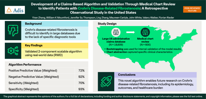
A validated claims-based algorithm could enable larger, more efficient studies of fibrostenosis in Crohn’s disease using real-world data — helping researchers and clinicians better measure how often stricturing...
AI models that combine CTE/MRE imaging with clinical and molecular data could improve objective classification of small-bowel Crohn's disease and help predict which patients are likely to respond to medical therapy...