Multimodal CCA + joint ICA reveals fused gray matter volume and resting-state functional alterations with neurotransmitter associations in Crohn’s disease
This study links brain structure and function differences to clinical measures and neurotransmitter systems in Crohn’s disease, which may help explain cognitive, mood, or quality-of-life effects some people with CD experience.
Who Should Pay Attention
Researchers and clinicians studying gut–brain interactions in IBD, and patients interested in how Crohn’s disease may relate to brain function, mood, or quality of life.
Study Snapshot
Story typeResearch paper
Evidence typeResearch paper
Source depthJournal abstract
What To Know
This journal abstract reports a multimodal neuroimaging study comparing 100 Crohn’s disease patients with 50 healthy controls using structural MRI (gray matter volume) and resting-state functional MRI (fALFF) analyzed by mCCA + jICA.
The authors identified joint and modality-specific brain components that differed between groups and that correlated with clinical measures (disease activity and quality of life).
The study also mapped those brain patterns to normative neurotransmitter distributions and found spatial correspondence with dopaminergic, serotonergic, noradrenergic, and opioid systems. The authors present these results as preliminary insights into gut–brain axis neurobiology in Crohn’s disease.
These findings come from the article abstract (structured-content depth: abstract) and summarize what the paper reports; they do not represent a clinical recommendation or proof of causal mechanisms.
Keep In Mind
the source provided is an abstract-level report of a clinical neuroimaging study. Results are preliminary and describe associations (not causal proof). Neurotransmitter mapping used normative distributions rather than direct patient neurotransmitter measurements.
Source Details
Review the original publication for the complete reporting, methods, and context.
This Cure8 brief is based on source text from the linked article. Cure8 is informational only and is not a substitute for professional medical advice, diagnosis, or treatment.
The study suggests Crohn’s disease is associated with coordinated brain structure and function differences that relate to disease activity and quality of life, pointing to brain–gut links that could inform future...
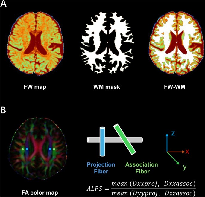
This study suggests brain glymphatic function — a fluid-clearance system — may be altered in people with Crohn’s disease and could relate to inflammation and common neuropsychiatric symptoms like fatigue, anxiety,...
If confirmed, a brain perivascular/glymphatic change linked to intestinal inflammation could help explain why sleep problems are common in Crohn's disease and point to new research targets for symptom mechanisms and...