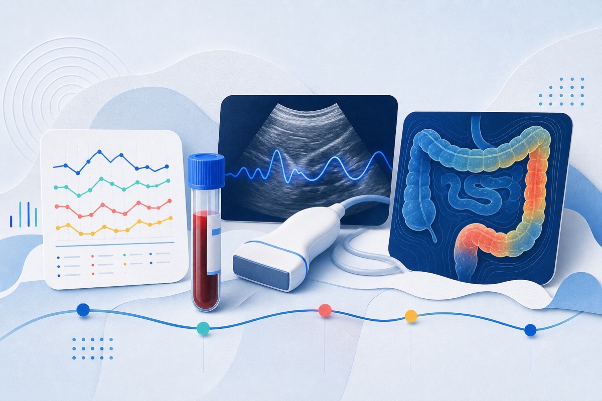
People with IBD have higher risk of several GI and hepatobiliary cancers; imaging is an important complement to endoscopic surveillance for detecting cancers that may be transmural or outside endoscopic reach.
Who Should Pay Attention
Clinicians (radiologists, gastroenterologists, surgeons), IBD multidisciplinary teams, researchers in imaging and IBD, and adult patients with long-standing IBD or concomitant primary sclerosing cholangitis.
Study Snapshot
Story typeResearch paper
Evidence typeResearch paper
Source depthJournal abstract
What To Know
This is an imaging-focused review summarizing typical and atypical radiologic features of malignancy across the small bowel, colon, rectum, perianal region, and hepatobiliary tree in IBD.
It highlights that malignant transformation often overlaps with chronic inflammatory changes and is best suspected when imaging behavior deviates from expected inflammatory patterns or shows interval progression.
The review emphasizes cross-sectional modalities (MRI, CT enterography), diffusion-weighted MRI, dual-energy CT, elastography, radiomics, and FAPI-based molecular imaging as emerging tools to improve detection and characterization. It also discusses common diagnostic pitfalls, mimics, and the value of structured reporting and multidisciplinary correlation.
Care implications Radiologists and IBD teams should compare current studies with prior imaging, look for discordant or progressive features, and escalate to endoscopy or biopsy when imaging raises concern. The article does not provide new treatment recommendations but frames imaging as a key component of cancer detection in IBD.
Keep In Mind
This entry is grounded in the journal abstract of a review article (Abdominal Radiology). It summarizes imaging considerations and emerging modalities but does not present original trial results. Clinical decisions should rely on multidisciplinary assessment and guideline-based surveillance.
Source Details
Review the original publication for the complete reporting, methods, and context.
Conflict statement: Declarations. Competing interests: The authors declare no competing interests. Third-party material: Regarding third-party material, we confirm that all necessary permissions for reproduced figures have been obtained. Permission for the figure reproduced from Neufert and Neurath ( Nature Reviews Gastroenterology & Hepatology, 2026 ) was obtained through the Copyright Clearance Center ( RightsLink ), and the license documentation is included with this submission. The figure reproduced from Faggiani et al. ( Cancers, 2024 ) is published under a Creative Commons Attribution ( CC BY 4.0 ) license, which permits reuse with appropriate attribution; the corresponding credit line has been included in the figure legend. Full attribution for both figures is provided in the manuscript.
This Cure8 brief is based on source text from the linked article. Cure8 is informational only and is not a substitute for professional medical advice, diagnosis, or treatment.
Perianal fistulas are a common and morbid complication of Crohn's disease in children; a reliable, less burdensome imaging tool could help assess severity and monitor response to therapy more often than MRI.
You can have active intestinal inflammation even without clear symptoms; early monitoring and tailored treatment aim to prevent complications and preserve quality of life.
Patients with Crohn's who develop intra-abdominal abscesses often face decisions about antibiotics, drainage, or surgery. Predictors like abscess size and imaging appearance may help estimate the chance of needing...